Duplex Study
What is Duplex Study?
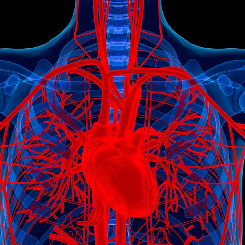
A carotid duplex scan is a simple and painless test that combines two types of ultrasound to check for blockages in your carotid arteries. Ultrasound is a type of scan that uses sound waves to create images inside your body. Your carotid arteries are located on both sides of your neck. Blocked carotid arteries are a major risk factor for stroke.
Duplex ultrasound uses high-frequency sound waves to observe blood flow velocity and the structure of leg veins. The term "duplex" means that two ultrasound methods are used: Doppler and B-mode. The B-mode transducer (similar to a microphone) produces an image of the examined vessel. The Doppler probe within the transducer assesses the speed and direction of blood flow in the vessel. For example, a carotid duplex scan may be performed to evaluate occlusion (blockage) or stenosis (narrowing) of the carotid artery and/or its branches in the neck. This type of Doppler exam provides a two-dimensional (2D) image of the artery, allowing determination of the artery’s structure and the presence of any material inside it, as well as the degree of blood flow.
")
Late Suresh Chandra Saha
Founder of J.D. PATHOLOGICAL LABORATORY CLINIC
")
Sibo Chandra Saha
Managing Director (MD)
")
Shaiba Saha Sree Krishna
Deputy Managing Director (DMD)
J D PATHOLOGY & CT SCAN CENTER
It has earned a strong reputation since its inception by ensuring accurate healthcare services.
| Saturday: 8 AM to 10 PM |
| Sunday: 8 AM to 10 PM |
| Monday: 8 AM to 10 PM |
| Tuesday: 8 AM to 10 PM |
| Wednesday: 8 AM to 10 PM |
| Thursday: 8 AM to 10 PM |
| Fridayday: 7 AM to 10 PM |